An asymptomatic wisdom tooth is not automatically healthy, but an impacted tooth is not automatically a surgical problem either. By comparing disease signs, tooth position, imaging, nerve risk and your ability to attend follow-up, you can identify when monitoring is safer and when removal offers the better balance of risks.
Key takeaways
- Monitor a disease-free wisdom tooth when it has no significant risk to nearby structures.
- Choose extraction for decay, infection, cysts, damage or a high-risk tooth position.
- Use panoramic X-rays to assess roots, bone, nerves and neighbouring teeth before deciding.
- Ask about surgical difficulty, recovery time and nerve-injury risk before removal.
When Is Monitoring Safer Than Removing a Wisdom Tooth?
Monitoring is safer than preventive removal when a wisdom tooth is disease-free and poses no significant risk to neighbouring structures. NICE recommends against prophylactic extraction of an asymptomatic, impacted tooth in that situation.
Evidence has not shown a clear overall benefit from routinely removing every asymptomatic, disease-free impacted wisdom tooth, although studies comparing removal with retention remain limited and low-certainty.
Monitoring suits a fully erupted tooth that is painless, free of decay, easy to clean, and surrounded by healthy gum and bone. It can also suit a partially or fully impacted tooth with no current pathology when you can attend scheduled reviews reliably.
Monitoring means active surveillance, not ignoring the tooth. Periodic examinations should check:
- Pain, swelling, drainage or restricted mouth opening
- Gum pockets and inflammation around the wisdom tooth
- Cavities in the wisdom tooth or second molar
- Periodontal damage and bone loss
- Resorption of the second molar
- Radiographic changes, including a developing cyst
One episode of pericoronitis does not automatically require extraction. Repeated episodes strengthen the case for removal because they show a recurring problem rather than an isolated event. Reconsider extraction if symptoms, decay, gum disease, bone damage, second-molar resorption or another abnormality develops.
Your age, medical status, tooth position and ability to maintain follow-up also affect whether surveillance remains the safer choice.
Which Findings Make Extraction the Better Choice?
Extraction becomes the better choice when a specific disease or structural threat is more dangerous to retain than the immediate risks of surgery.
| Finding | Why removal may be justified |
|---|---|
| Unrestorable decay | The tooth cannot be predictably repaired. |
| Untreatable pulp or root-tip disease | Root-canal treatment cannot control the disease. |
| Abscess or cellulitis | Infection is spreading or cannot be controlled locally. |
| Repeated pericoronitis | Recurrent gum infections create a stronger case than one isolated episode. |
| Gum disease | Bone loss or deep pockets affect the wisdom tooth or second molar. |
| Cyst or tumour | A lesion threatens surrounding bone or teeth. |
| Second-molar damage | Resorption, decay or another defect is caused or worsened by the wisdom tooth. |
| Fracture | The tooth cannot be restored reliably. |
| Planned treatment | The tooth obstructs jaw surgery or compromises orthodontic movement. |
A painless tooth can still be unhealthy. An X-ray may reveal hidden decay, bone loss, resorption or a cyst before you feel anything.
Impaction alone is not a diagnosis. A dentist may instead restore a repairable cavity, treat the adjacent tooth, clean inflamed gum or manage an infection before recommending removal. The decision should identify the actual disease, whether treatment can save the tooth, and what is likely to happen if you wait.
Extraction is justified when that expected harm outweighs the surgical risk.
How Do Position and X-Rays Change the Decision?
Position and X-rays help determine whether an impacted wisdom tooth needs surveillance, straightforward removal, surgical removal or a nerve-sparing alternative.
- Mesioangular: points toward the second molar.
- Vertical: follows the normal tooth axis.
- Horizontal: lies on its side.
- Distoangular: tilts away from the second molar.
The examination checks whether the tooth has erupted, how much gum covers it, whether you can clean around it, and whether it causes tenderness, deep gum pockets or decay. The clinician also assesses mouth opening, because limited access can turn an apparently simple extraction into surgery.
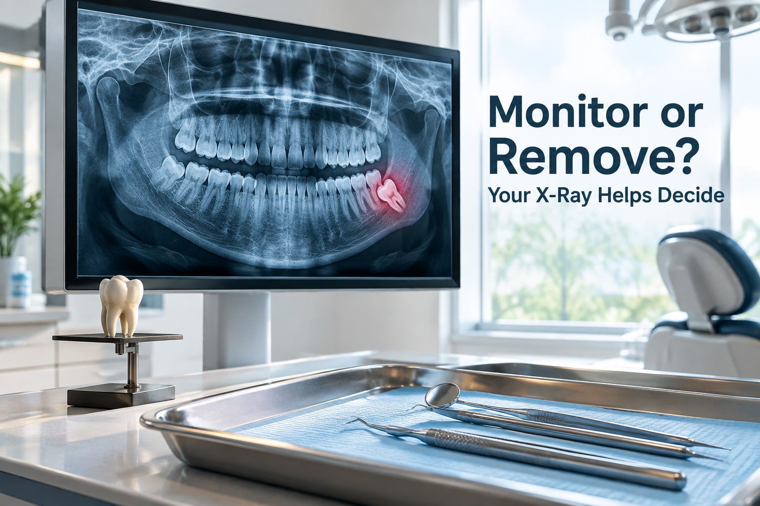
A panoramic radiograph commonly shows the wisdom tooth, its roots, the second molar, surrounding bone and the lower jaw nerve canal. It helps reveal hidden decay, bone loss, root shape and the tooth’s angulation.
CBCT is not routine for every patient. It is most useful when three-dimensional detail about the inferior alveolar nerve, lingual area, roots or another structure could change the plan between monitoring, complete extraction and coronectomy.
For a lower tooth whose roots closely contact the inferior alveolar nerve, coronectomy may suit selected cases. The clinician removes the crown but leaves the roots, reducing direct nerve-injury risk; root migration or later symptoms can require another operation. This is one form of impacted wisdom tooth treatment, not an automatic mandate for extraction.
What Are the Wisdom Tooth Extraction Options and Their Risks?
Removing a diseased or high-risk wisdom tooth reduces the chance of future decay, infection, cyst formation, gum damage or injury to the second molar. You exchange that possible future harm for immediate surgical risks.
- Monitoring means scheduled examinations and X-rays rather than ignoring the tooth. It suits a painless tooth with no current disease when follow-up is reliable.
- Conventional extraction removes a fully erupted tooth when enough of its crown is accessible for dental instruments.
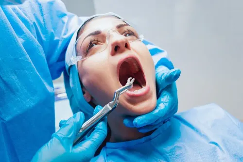
- Surgical extraction is used when gum or bone blocks access. It can involve a gum incision, removal of a small amount of bone and removal of the tooth as one piece.
- Sectioning divides the tooth into smaller pieces before removal, reducing the force needed when roots or position make one-piece removal difficult.
- Selected coronectomy removes the crown of a lower wisdom tooth but leaves its roots when they lie close to the inferior alveolar nerve. Root migration or later symptoms can require another operation.
The risks extend beyond expected soreness: dry socket, infection, bleeding, swelling, restricted mouth opening and damage to the adjacent second molar can occur. Injury to the inferior alveolar or lingual nerve can cause temporary or permanent altered sensation.
Nerve risk depends on tooth position and nerve proximity, not on whether the procedure is described as comfortable. Extraction is not risk-free prevention, and monitoring is not risk-free neglect; the choice balances surgical harm now against disease and structural damage later.
How Should You Choose Between Monitoring and Removal Locally?
Choose the plan that explains the tooth’s actual disease risk, imaging and follow-up—not the listing you found for wisdom tooth extraction in Hinjewadi, wisdom tooth removal Wakad or an oral surgeon near Hinjewadi.
Ask these four questions before agreeing:
- What disease or future structural risk has been identified, and what could happen if treatment waits?
- Is the tooth fully erupted, partially impacted or classified by a specific angulation, such as mesioangular, vertical, horizontal or distoangular?
- Does the panoramic image show proximity to the inferior alveolar nerve, and would a CBCT scan change the choice between monitoring, full removal and coronectomy?
- What are the alternatives, anaesthesia plan, expected recovery, warning signs and follow-up schedule?
| Option | What it means | When it fits |
|---|---|---|
| Monitoring | Scheduled examinations, symptom checks and repeat imaging when needed | No disease, manageable risk and reliable reviews |
| Removal | Conventional or surgical extraction, with sectioning or bone removal when required | Disease, damage or a future risk outweighs surgical risk |
| Coronectomy | Crown removal while selected roots remain | Lower tooth with close nerve contact when full extraction carries greater nerve risk |
Your age, medical conditions, pregnancy planning, smoking, oral hygiene and ability to attend reviews change the balance. Ask how each affects your specific case.
A consultation at Deshmukh Dental Clinic can help compare examination findings, imaging, monitoring and referral or removal in the Hinjewadi and Wakad area. Judge the explanation and follow-up plan, not location alone.
Related service
Oral Surgery Wisdom teeth, no problem! 😊 Our expert team handles wisdom tooth extractions with care and precision, ensuring a smooth and comfortable experience. View service → |
Frequently asked questions
When is monitoring safer than removing a wisdom tooth?
Monitoring is safer when the tooth is disease-free, symptom-free and does not threaten neighbouring teeth, bone or nerves. Arrange regular dental reviews and repeat imaging when your dentist recommends it.
Which findings make wisdom tooth extraction the better choice?
Extraction is usually the better choice when there is repeated infection, decay that cannot be restored, gum disease, a cyst or tumour, damage to the second molar, or another clear disease risk.
How do position and X-rays change the wisdom tooth decision?
A panoramic X-ray shows the tooth angle, eruption space, roots, bone and relationship to the inferior alveolar nerve. Your dentist may request a CBCT scan when a three-dimensional view is needed for surgical planning.
What wisdom tooth extraction options are available?
Options include monitoring, simple extraction for an erupted tooth, and surgical removal for an impacted tooth. Risks include pain, swelling, infection, dry socket and, for lower teeth near the nerve, temporary or permanent altered sensation.
How should you choose between monitoring and removal locally?
Compare the examination findings, X-ray evidence, surgical complexity, recovery plan and nerve risk. For wisdom tooth extraction in Hinjewadi or wisdom tooth removal in Wakad, ask whether the dentist or oral surgeon can manage your specific tooth position.
Keywords